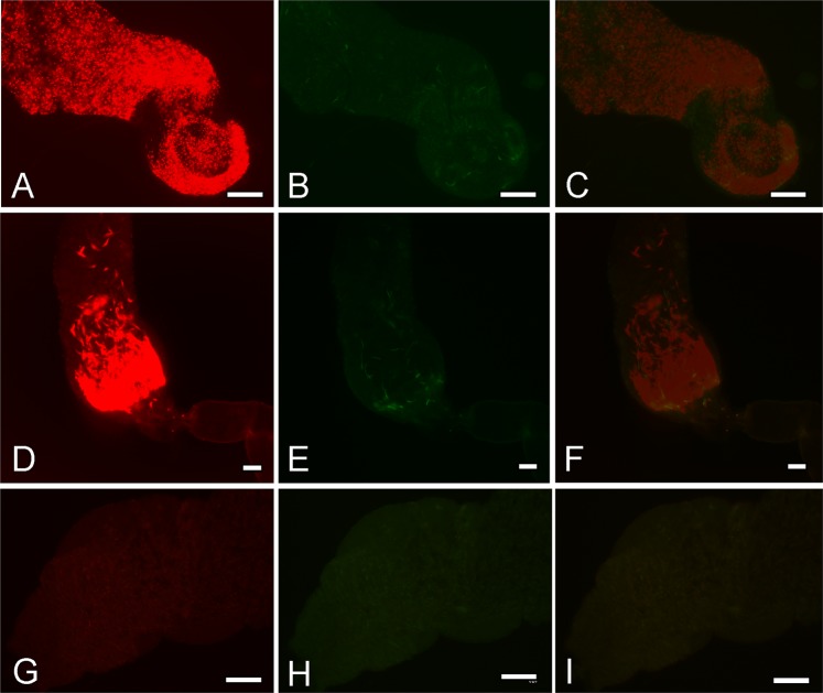
Figure 2.
Fluorescence micrographs of thoracic midguts with cardia section and stomodeal valve of Lutzomyia spp. females on days 8 postinfection. Cardia of Lu. longipalpis (A–C) and Lu. migonei (D–F) coinfected by Leishmania (Viannia) braziliensis (green) and Leishmania (Leishmania) infantum (red). Control uninfected gut of Lu. longipalpis (G–I). (A,D,G) images from red fluorescence, (B,E,H) images from green fluorescence and (C,F,I) merged images, scale bar: 5 mm.

