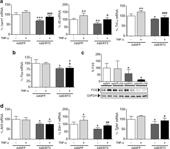
Fig. 4.
SIRT3 modulates inflammation and FOS levels in neonatal rat cardiomyocytes. Relative quantification of Icam1, Il6, Tnf-α a, Fos b, Atf4, Edn-1, and Tgfb1 d mRNA expression in neonatal rat cardiomyocytes overexpressing Sirt3 (AdSIRT3) or GFP control vector (AdGFP; 30 IFU per cell, for 48 h) in the presence or absence of TNF-α (TNF, 10 ng/mL, 24 h). The graphs represent the quantification of the Gapdh–normalized mRNA levels expressed as a percentage of the control samples ± SD. c Western blot analysis showing FOS protein levels in total protein extracts obtained from the same samples depicted in panel a. The graph represents the quantification of protein levels normalized to GAPDH expressed as a percentage of the control samples ± SD. The data were compared by ANOVA followed by Tukey’s post hoc test. *P < 0.05, **P < 0.01, and ***P < 0.001 vs. AdGFP; #P < 0.05, ##P < 0.01, and ###P < 0.001 vs. AdGFP + TNF-α

