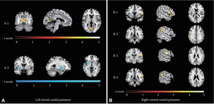
Figure 1.
t-statistic maps of the significant FC of the left DCP (A) and right VRP (B) in patients with OCD compared to healthy controls. The FC between the left DCP and the right intracalcarine cortex was stronger in patients with OCD than in healthy controls (red through yellow; A-1), whereas the opposite was true of the FC between the left DCP and the left putamen (shades of blue; A-2). The FC values between the right VRP and the right inferior frontal gyrus (B-1), left supramarginal gyrus (B-2), right postcentral gyrus (B-3) and right supramarginal/angular gyrus (B-4) were stronger in patients with OCD (red through yellow). All clusters survived false discovery rate and Bonferroni correction, with p-values<0.05/12. FC: functional connectivity, DCP: dorsal caudal putamen, VRP: ventral rostral putamen, OCD: obsessivecompulsive disorder.

