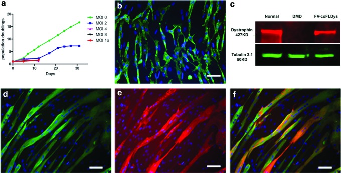
Figure 2.
In vitro transduction of human DMD myoblasts with FV-coFLDys. (a) In vitro proliferation curve of cells transduced with different amounts of FVs, for 30 days after their transduction. (b) The expression of dystrophin in cells with FV of MOI 2. (c) Western blot indicating the correct molecular weight of the transgene in differentiated myotubes derived from the transduced cells. (d–f) Immunostaining of myosin heavy chain (d, green), dystrophin (e, red), and merged images (f). Nuclei were counterstained with DAPI. Scale bar = 25 μm. coFLDys, codon-optimized full-length dystrophin; DMD, Duchenne muscular dystrophy. Color images are available online.

