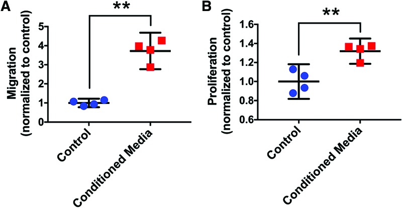
Figure 1.
Migration (A) and proliferation (B) of HFF1 dermal fibroblasts both increased after exposure to MSTC-conditioned media for 24 h, compared with control (unsupplemented) media. Mean ± 95% CI. **p < 0.005, n = 4. CI, confidence interval; HFF1, human foreskin fibroblasts; MSTC, micro skin tissue column. Color images are available online.

