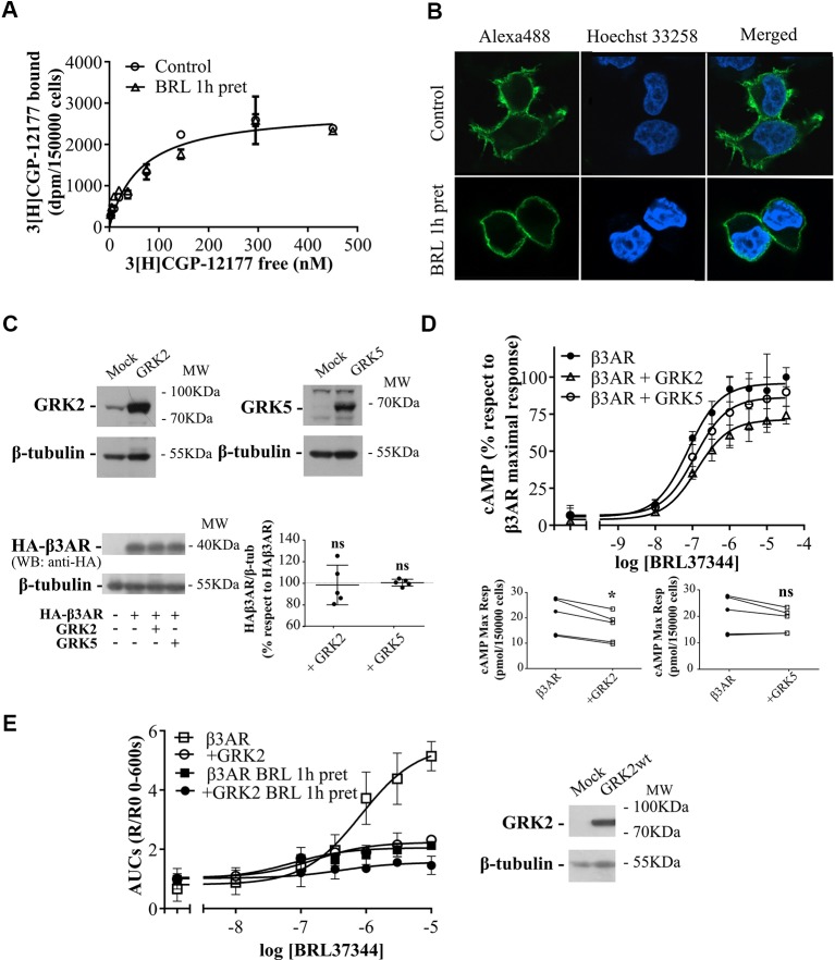
Figure 3.
Mechanisms of regulation of β3AR cAMP response. (A) HEK293T-β3AR cells were pre-treated with 10µM BRL37344 for 1h or not, washed and incubated with different concentrations of 3[H]-CGP12177 as stated in Materials and Methods. Data represent mean ± SD (n=3). Best-fit values of the parameters are in the main text. Comparisons of Bmax from both binding curves were done by extra sum of squares F-test. (B) HEK293T cells transfected with HAβ3AR were pre-treated with 10µM BRL37344 for 1h or not, washed and fixed for immunocytochemical staining as described under Materials and Methods. Images of fluorescence signal corresponding to secondary Alexa488-conjugated antibody, nuclear Hoechst 33258 staining and merged images are shown. The immunostaining obtained with the anti-HA tag antibody indicated expression and location of HAβ3AR in HEK293T cells after transfection. Images are representative of 3 independent experiments. (C) HEK293T-β3AR cells co-transfected with GRK2 or GRK5 were lysed, resolved by 12% SDS-PAGE and immunoblotted using anti-GRK2, anti-GRK5, anti-HA or anti-β tubulin antibodies. A representative image is showed. Densitometry analysis was performed with ImageJ as indicated in Materials and Methods and analyzed by student t-test comparing HA-β3AR levels in GRK2 or GRK5 co-transfected cells vs HA-β3AR without GRKs which was considered as 100%. Results are mean ± SD (n=5). (D) HEK293T-β3AR cells co-transfected with GRK2 or GRK5, were pre-treated with IBMX for 3 min and stimulated with different concentrations of BRL37344 for 30 min. Upper panel: results are expressed as % respect to maximal response of control (β3AR without GRKs), data represent mean ± SD (n=5). Lower panels: Raw data of the maximal cAMP levels from each individual experiment are shown. Data were analyzed by paired student t-test. ns, not significant; *P < 0.05 respect to β3AR. cAMP production was quantified as described under Materials and Methods. (E) HEKT Epac-SH187 transiently co-transfected with β3AR and GRK2 wild type were pre-treated during 1 h with either 10µM BRL37344 or vehicle. After that, cells were washed and challenged with different concentrations of BRL37344. Concentration-response curves were constructed with the AUC values of 10-minute R/R0 i-cAMP response of time course of FRET changes determined in FlexStation® 3 at 37°C as described in Materials and Methods (left). HEKT Epac-SH187 co-transfected with β3AR and GRK2 wild type, were lysed, resolved by SDS-PAGE 12% and immunoblotted using anti GRK2 or anti-β tubulin antibodies as indicated. Image showed is representative of three independent experiments (right).

