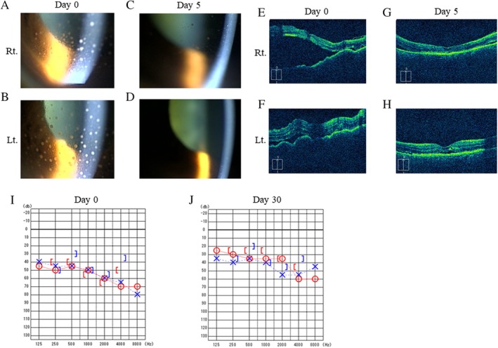
Figure 2.

Anterior segment photography before pulse corticosteroid therapy (A, B) and after five days (C, D). Mutton‐fat keratic precipitates on the corneal endothelium were improved after steroid therapy. Optical coherence tomography images before pulse corticosteroid therapy (E, F) and after five days (G, H). Bilateral retinal detachment showed significant improvement after steroid therapy. Audiograms obtained before treatment (I) and 30 days after steroid therapy (J). Bilateral auditory acuity showed improvement after steroid therapy.
