Figure 1.

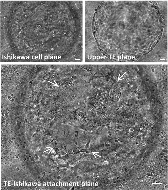
Phase contrast image of a live day 8, human blastocyst attached to Ishikawa cells after 48 h in co-culture. An area of trophectoderm (TE)-mediated attachment to Ishikawa cells (arrows) is visible. Scale bars 20 μm.

Phase contrast image of a live day 8, human blastocyst attached to Ishikawa cells after 48 h in co-culture. An area of trophectoderm (TE)-mediated attachment to Ishikawa cells (arrows) is visible. Scale bars 20 μm.