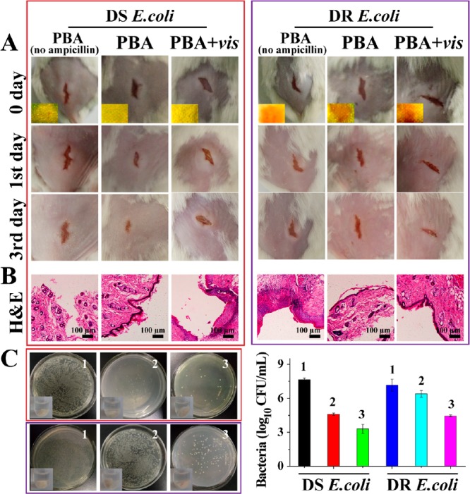
Figure 5.

Use of PBA for wound disinfection in mice. (A) Photographs of wounds on the mice. Inset images in the first row revealed the color on PBA. (B) Photomicrographs showing section of skin tissues with H&E staining. (C) Bacteria was separated from wound tissue and then cultured on agar plates. The upper row belongs to DS E. coli, and the lower row belongs to DR E. coli. 1, PBAno ampicillin; 2, PBA; 3, PBA+vis.
