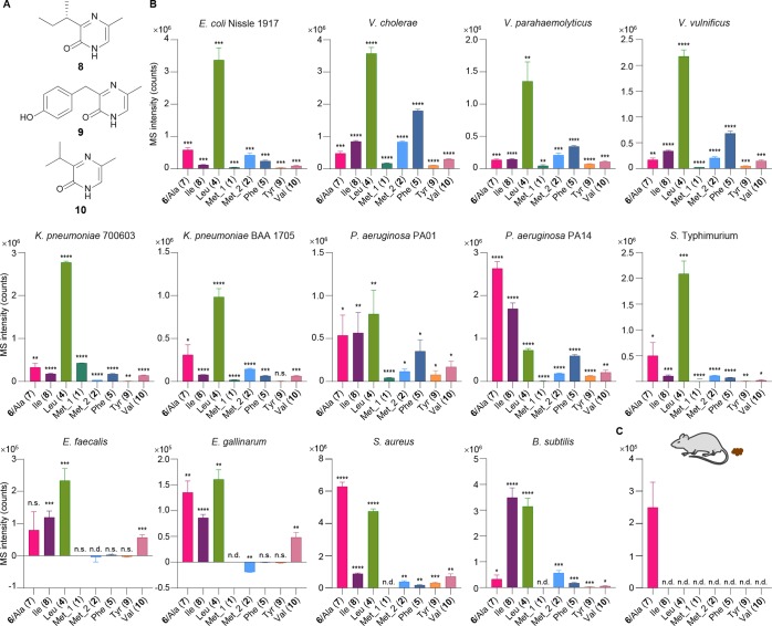
Figure 6.
Pyrazinone profiling in different bacterial strains. (A) Structures of pyrazinones derived from l-Ile (8), l-Tyr (9), and l-Val (10). (B) Production of the pyrazinones derived from l-Thr (right-hand side) and l-Ala (7, minor in E. coli, which is indistinguishable with major 6), l-Ile (8), l-Leu (4), l-Met (2), l-Phe (5), l-Tyr (9), or l-Val (10) (left-hand side), and 3R-hydroxy-l-Leu (right-hand side) and l-Met (1) (left-hand side) from E. coli Nissle 1917, V. cholerae, V. parahaemolyticus, V. vulnificus, K. pneumonia 700603, K. pneumonia BAA 1705, P. aeruginosa PA01, P. aeruginosa PA14, S. enterica serovar Typhimurium, E. faecalis, E. gallinarum, S. aureus, and B. subtilis. Compounds 6 and 7 are indistinguishable in these studies (Figure S2C). Data are presented by mean ± SD of MS intensities (counts) observed in bacterial culture extracts subtracted by the mean of the media background. Statistical analysis was performed between the bacterial culture extracts (n = 3) and media control extracts (n = 3) using two-tailed t-test. *P < 0.05, **P < 0.01, ***P < 0.001, ****P < 0.0001; ns, not significant; nd, not detected. (C) A major peak corresponding to pyrazinones 6/7 was detected in C57BL/6 mouse fecal samples. n = 5 biological replicates. Data are mean ± SD.

