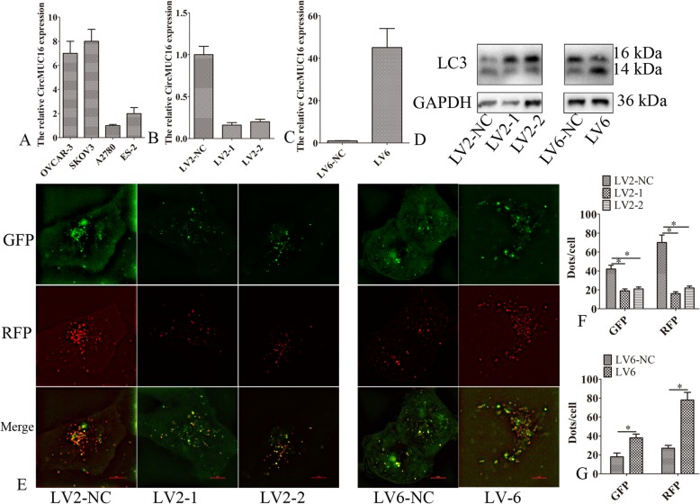
Fig. 2.
CircMUC16-mediated autophagy flux of epithelial ovarian cancer. a CircMUC16 expression was determined in four cell lines by qPCR. b CircMUC16 expression was detected in SKOV3 cells. c The expression of circMUC16 was detected in A2780 cells. d Western blot was used to detect the expression of LC3-II after silencing or ectopic expression of circMUC16. e mRFP-GFP-LC3 distributions in SKOV3 and A2780 cells were analyzed by confocal microscopy after silencing or ectopic expression of circMUC16. The symbols * and ** show p < 0.05 and 0.01, respectively. Scale bar: 5 μm

