Figure 7.
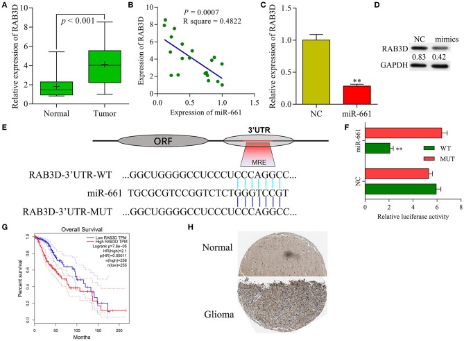
RAB3D served as a target of miR-661. (A) RAB3D mRNA expression in samples of normal and tumor tissues was examined by qRT-PCR. (B) Person's correlation coefficient was used to analyze the correlation between RAB3D and miR-661. (C,D) RAB3D expression was measured by qRT-PCR and western blot assays after transfection with miR-661 mimics (**P < 0.01). (E) The binding site between RAB3D and miR-661 was identified. (F) 293T cells were co-transfected with WT-RAB3D or MUT-RAB3D and miR-661 or NC; dual luciferase reporter assays were used to analyze relative luciferase activity. (G) GEPIA showing the overall survival of glioma patients. (H) RAB3D expression in glioma and adjacent non-tumor tissues was assessed by immunochemistry (online database: The human protein atlas).

