
Figure 1.
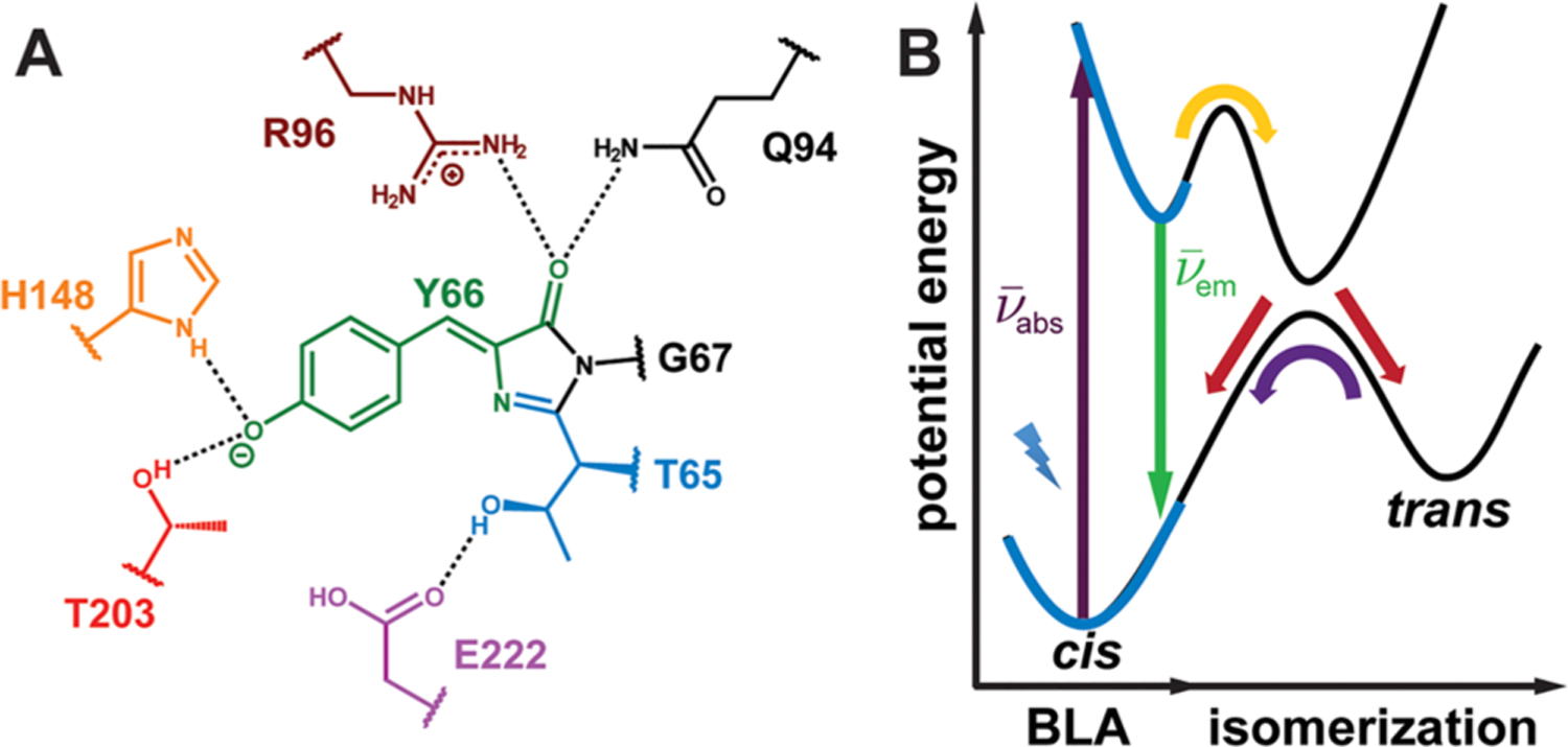
Structure and energetics of the GFP chromophore embedded in the protein environment. (A) The GFP chromophore, 4-hydroxybenzylideneimidazolinone (HBI), and surrounding residues within GFP. The colored residues represent those mutated in this work (Table 1). (B) Potential energy diagram of the ground and excited electronic states for the GFP chromophore.14 In this work, we focus on the region of the diagram highlighted by the blue boldface curves associated with absorption () and emission (). The nuclear motion relevant to this part of the potential energy diagram is the bond length alternation (BLA).