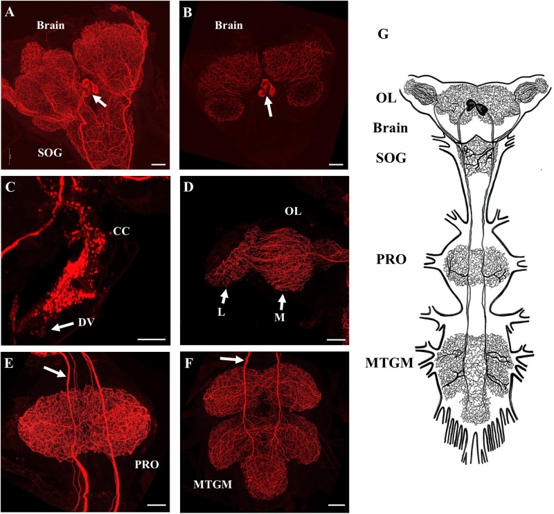
FIGURE 2.
SIFamide-like immunoreactivity (SLI) in the central nervous system (CNS) of fifth instar R. prolixus. The staining pattern seen here was the same for male and female fifth instar R. prolixus. (A) Ventral view of the brain and the sub-esophageal ganglion (SOG), cell bodies indicated by arrow. (B) Dorsal view of the brain showing the two pairs of cell bodies (arrow). (C) SLI in processes in the corpus cardiacum (CC) extending onto the dorsal vessel (DV). (D) Projections from the SIFamidergic neurons extending into the optic lobe (OL), medulla (M), and lamina (L). (E) View of the prothoracic ganglion (PRO) with axons continuing from the SOG. The arrow points to separations in the axons that exist as closely oriented pairs. (F) View of the mesothoracic ganglionic mass (MTGM) with axons (arrow) continuing from the PRO. (G) Diagrammatic representation of the two pairs of cell bodies and their extreme projections throughout all ganglia. Map is drawn from over 20 preparations. Scale bars = 50 μm.

