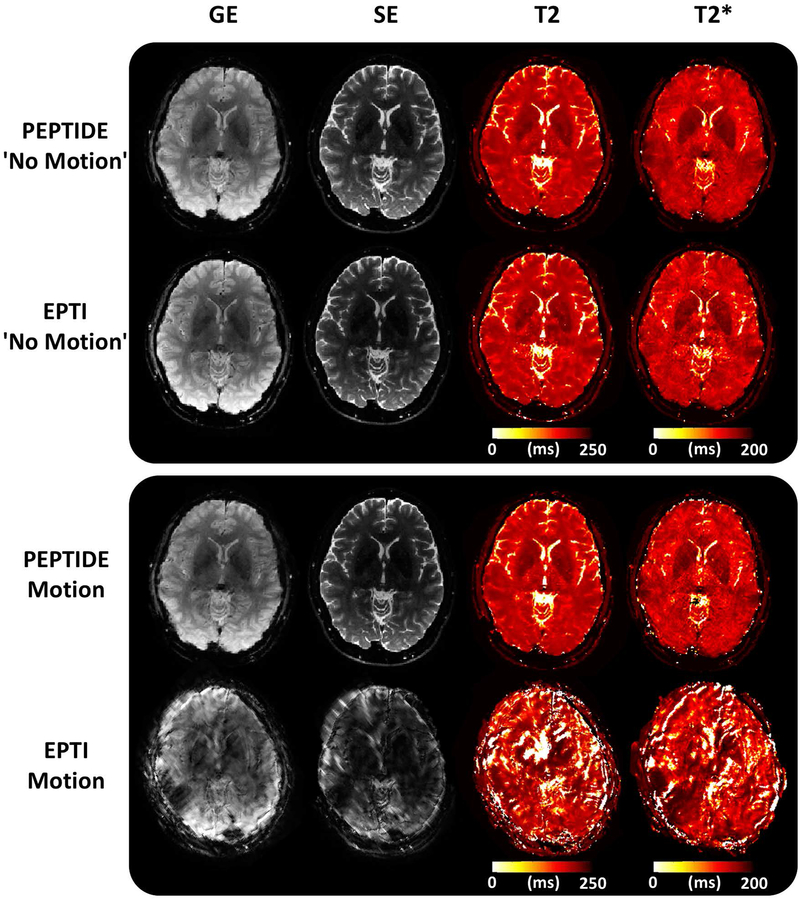
Figure 7 –
PEPTIDE and EPTI comparison. A single image from the gradient-echo (GE) and spin-echo (SE) acquired time series and calculated T2 and T2* maps are shown in a case without motion for PEPTIDE and EPTI, as well as for a case with identical severe (~20° rotation) motion for both - corresponding to the red color-coded motion from Figure 7. Both methods were reconstructed with the same number of blades/segments = 7.

