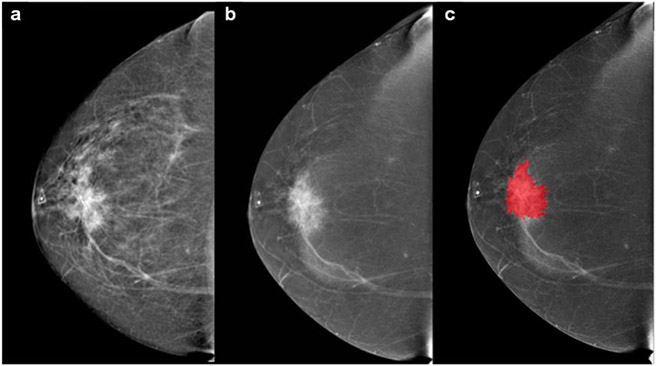
Fig. 2.
55-year-old patient with a palpable mass in the right breast. a Cranio-caudal full-field digital mammography of the right side. Scattered fibroglandular breasts. Corresponding to the palpable finding (triangular marker) at 12:00 in the anterior depth is an irregular shaped, spiculated 4 cm mass that is highly suspicious for malignancy. b Cranio-caudal contrast enhanced mammography. The examination was performed using full breast digital technique after injection of 153 ml of Omnipaque 350. Mild background parenchymal enhancement. The palpable suspicious mass enhances with contrast administration. c The lesions borders were segmented with the MAZDA segmentation tool to run the radiomics analysis. BI-RADS 5: Highly suspicious lesion and biopsy is recommended. The final histology yielded invasive ductal carcinoma (HR positive, HER2 negative, grade 2)

