
Figure 1.
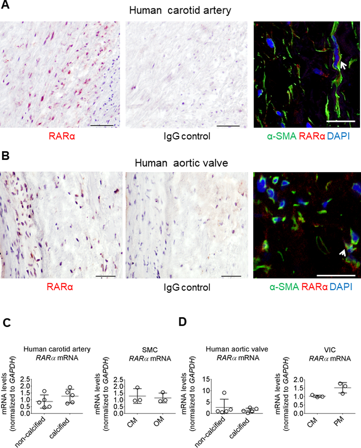
Human cardiovascular tissue contained immunoreactive retinoic acid receptor. (A) Human carotid artery and (B) aortic valve RARα immunohistochemistry (red color); scale bars, 50 μm. IgG control antibody immunohistochemistry staining for matching donor adjacent sections used as negative controls. RARα (red color) immunofluorescence showing localization with α-smooth muscle actin (α-SMA) positive cells (green color) in human carotid artery (white arrow) and with vimentin-positive cells (green color) in human aortic valve (white arrow); scale bars, 20 μm. One of five carotid artery and aortic valve donors shown, with representative RARα immunohistochemistry shown for two additional donors in Figures I and II in the online-only Data Supplement. (C) Human carotid artery, SMC, (D) aortic valve, and VIC RARα mRNA levels in calcified and non-calcified tissues (n = 5 donors) and CM, OM or PM treated cells after two weeks in culture (n = 3 donors). Error bars indicate STDEV of mean. Data analyzed by Student’s t-test.