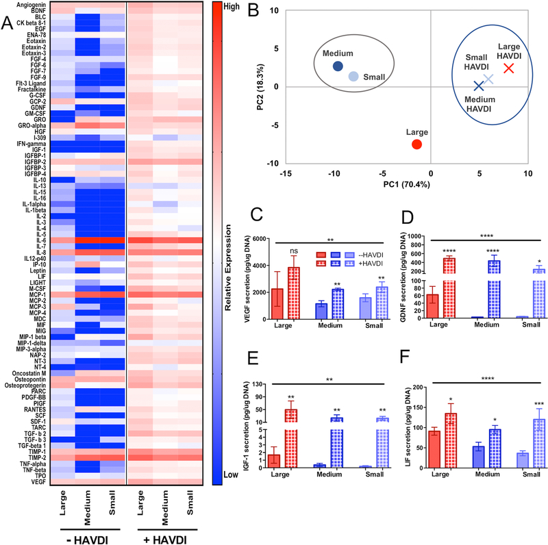
Figure 6 – HAVDI inclusion in microgel scaffolds increases secretory phenotype of hMSCs.
(A) Heatmap of cytokine expression of encapsulated hMSCs in large (190±100μm), medium (110±60μm), and small (13±6μm) diameter microgel scaffolds with and without inclusion of the HAVDI peptide. (B) PCA analysis of hMSC secretory profile between large (red), medium (blue), and small (light blue) microgel scaffolds without (circles) and with HAVDI (X symbol). PC1 and PC2 explained 70.4% and 18.3% of the variance respectively. (C) ELISA quantification of VEGF, (D) GDNF, and (E) IGF-1. HAVDI conditions are represented by hashed bars. Stars represent significance relative to respective unmodified scaffolds. Overall significance (top bar) determined using a one-way ANOVA with multiple comparisons. ****p<0.0001, ** p<0.01, n.s. – non-significant

