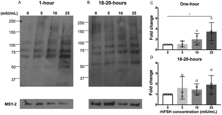
Figure 1. Rec. hFSH treatment induces phosphorylated PKA substrates in a dose-dependent manner in secondary follicles.
(A, B) Immunoblot analysis of phosphorylated PKA substrates in murine follicles following rec. hFSH treatment for one-hour (A) and 18-20 hours (B). Densitometry analysis of phospho-PKA substrate protein levels were performed and values indicated (C, D) were normalized to MSY-2 levels. The experiment was repeated 5 times (3 times for 5mIU/mL concentration) with 10 follicles in each group. Representative images are shown. The arrow indicates the phospho-PKA substrate band that was chosen for densitometry. Data are expressed as mean ± S.D. * P<0.05.

