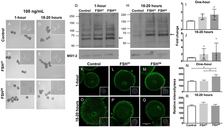
Figure 3. Treatment of follicles with 100ng/mL individual purified FSH glycoforms for one-hour results in differential regulation of phospho-PKA substrates.
(A-F) Follicles were morphologically intact with healthy oocytes following treatment with 100 ng/mL FSH glycoforms at the one-hour and 18-20-hour time points. Scale bar = 400μm (100μm for insets). (G, H) Immunoblot analysis of phosphorylated PKA substrates in murine follicles following FSH glycoform treatment for one-hour (G) and 18-20 hours (H). (I, J) Densitometry analysis of phospho-PKA substrate protein levels were performed and values were normalized to MSY-2 levels. Experiment was repeated 4 times with 10 follicles per group. (K-R) Immunofluorescence analysis of phosphorylated PKA substrates in murine follicles following FSH glycoform treatment for one-hour (K-M) and 18-20 hours (O-Q). Single optical sections are shown. Insets show DNA in blue and F-actin in red. Images corrected to +20% brightness and −20% contrast. (N, R) Relative intensity per area of all follicles examined is plotted for each time point. Between 13-20 follicles were examined at each time point. Data expressed as mean ± S.D. Scale bar = 50μm. * P<0.05.

