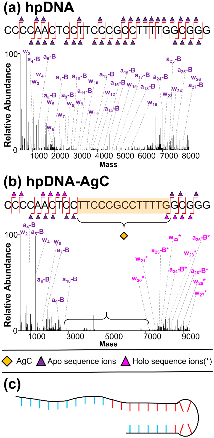
Figure 4.

Deconvoluted a-EPD spectra and sequence coverage maps of 6- hpDNA CCCCAACTCCTTCCCGCCTTTTGGCGGG (a) without and (b) with AgC, highlighting localization of the AgC, (c) model of hpDNA-AgC with AgC-coordinating nucleobases in red and noncoordinating nucleobases in blue. Sequence ion assignments from deconvoluted a-EPD spectra are summarized in Figures S25 and S26.
