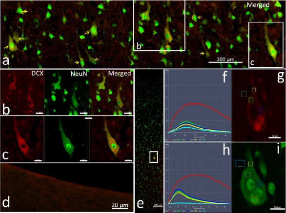
Figure 3.

Antigen adsorption of DCX antibodies.
Antibody combination of the antigen-adsorbed DCX antibody ab18723-1 and the secondary antibody ab150072 (unadsorbed) was first used to label the adjacent sections of the cortex and subventricular zone. Panel a is taken from the fifth layer of the cortex (Additional Figure 4 (527.9KB, tif) ) to show the NeuN/DCX double-labeled neurons (yellow arrows). In b and c image series from the white boxes in panel a, DCX immunoreactivity (red) was reduced after adsorption, but medium intensity red fluorescence persisted. Panel d shows no DCX-positive labeling in the subventricular zone after the DCX antibody was adsorbed by DCX antigen. To distinguish the source of red fluorescence that remains after DCX antibody adsorption, the adjacent sections of adult macaque cortex were immunolabeled with the antigen-adsorbed DCX antibody (ab18723-1) and the secondary antibody (ab150072) that had been pre-adsorbed with a rat cortex section. Panel e shows the localization of the tested neuron in the cortex (white box). Panels f and h show the spectral features of the images in g and i, respectively. In graph g and i, small boxes in different colors represent the locations of spectral analysis spots. Red: Autofluorescence; green, blue and yellow represent immunofluorescence from the tested sites; light blue: the background. In these sections labeled by antigen pre-adsorbed DCX antibody, the immunofluorescence intensity of some neurons is still higher than the background, indicating the presence of cross reactivity. Scale bars: 200 µm in panel e, 100 µm in panel a, and 20 µm in panels b–d, g and i. DCX: Doublecortin; Lv: lateral ventricle.
