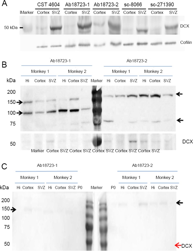
Figure 6.

Comparison of western blot assays of the adult macaque brain using the various antibodies.
(A) Differences in western blot results among the various DCX antibodies. Either cortex or SVZ of the same monkey was lysed, and 25 μg of total protein was loaded per lane. Polyvinylidene fluoride membranes were incubated with different DCX antibodies and were aligned for image development. Cofilin was used as control. (B) Western blot showing the non-specificity of DCX antibodies. Different batches of Ab18723 were used to detect the target protein from monkeys. A total of 25 μg protein from the hippocampus, cortex and SVZ of rhesus macaques were loaded per lane. The blot was cut for antibody incubation and the strips were aligned before image development. Protein bands not mentioned in the antibody datasheet were observed in monkeys but not in rats. The molecular weight of those bands differed between batches. (C) The non-specific band could not be completely blocked by pre-adsorption. Tissue pre-adsorbed DCX antibodies Ab18723-1 and 2 were used to detect DCX in 6 μg total brain protein from P0 rat and 25 μg total protein from the hippocampus, cortex and SVZ of adult rhesus macaques. The predicted DCX band (40–45 kDa) disappears in every lane, while the non-specific band is attenuated but not completely eliminated. Arrows on either side indicate the molecular weights. CTS: Cell Signaling Technology; DCX: doublecortin; Hi: hippocampus; SVZ: subventricular zone.
