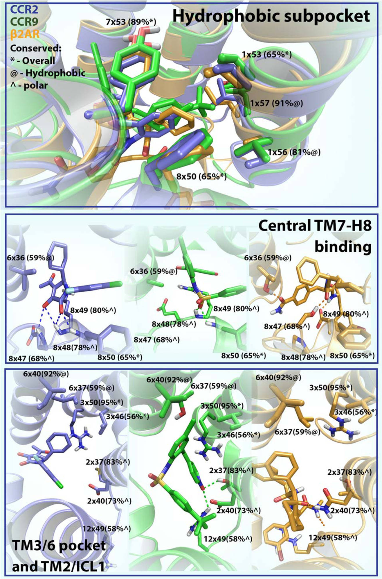
Figure 3. Overview of structural features of the intracellular binding site.

Common features in intracellular ligand binding derived from the crystal structures of CCR2 (PDB 5T1A), CCR9 (PDB 5LWE) and β2AR (PDB 4XT1). Residues are numbered using structure-based Ballesteros-Weinstein numbers [15]. Residue conservation among all class A GPCRs is shown in the following way; residues that are overall conserved (identical) in class A (>50%) are shown first (*); for residues that are not conserved we show how conserved they are in terms of polarity (^) or hydrophobicity (@). The three different boxes represent three different sections of the intracellular binding sites, in the upper panel all receptors are superimposed while in the lower two boxes the receptors are shown separately. CCR2 is colored blue, CCR9 is colored green and β2AR is colored orange.
