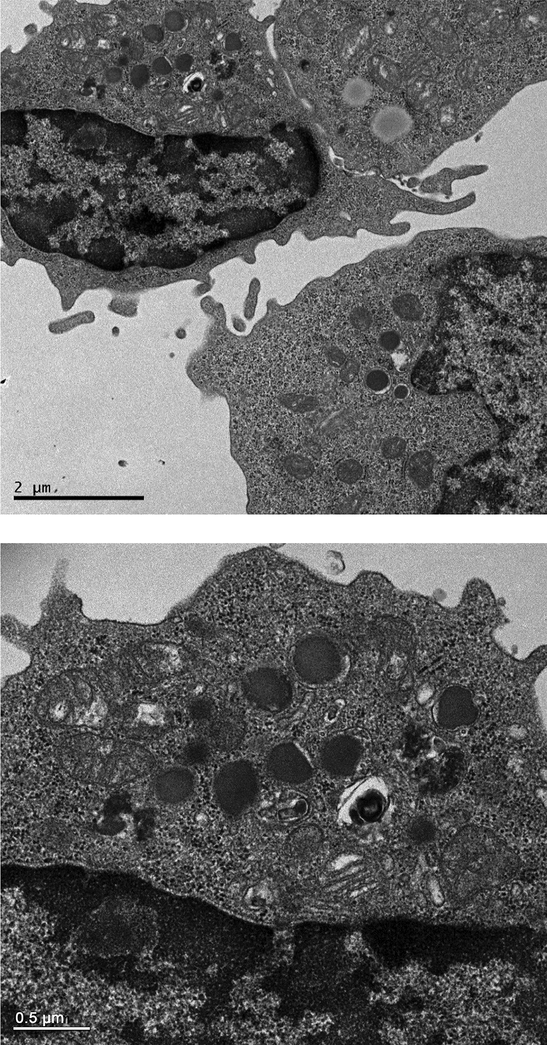
Figure 8. Transmission electron micrographs of CD94 selected cells.

Electron-dense granules and reniform nuclei, typical of LGL, are shown in both figures. Top photo is 2000X and the lower is 5000X magnification with the bars indicating 2.0 and 0.5 microns, respectively.
