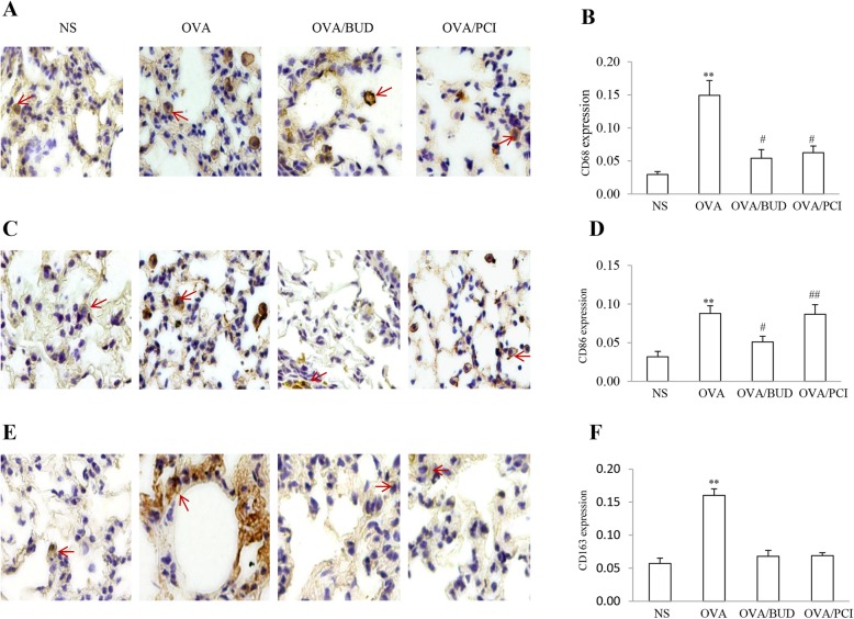
Fig. 3.
Macrophage polarization states. Distribution and proportion of CD68+ (a), CD86+ (c) and CD163+ (e) macrophages (brown) were identified using IHC procedures. Intensity for the color response to antibodies was visible in lung tissues from all groups. Population proportions for the stained cells as shown by red arrows were calculated as a fold change of the control. (b, d, f). The results were expressed as Mean ± SD (n = 3). **: a p-value of < 0.01 vs NS, OVA/BUD and OVA/PCI except the number of CD86+ cells in OVA/PCI group. #: P < 0.05 vs NS and ##; P < 0.01 vs either NS or OVA/BUD group

