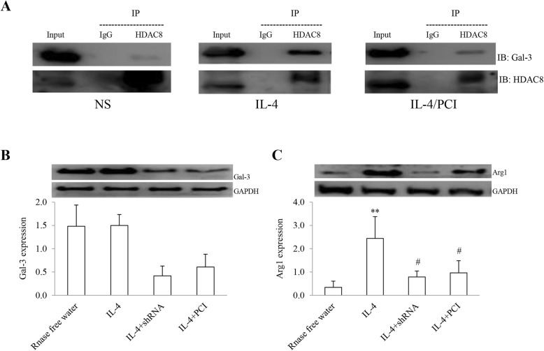
Fig. 7.
Identification of HDAC8 and Gal-3 interaction. HDAC8 and Gal-3 (a) in protein complex from cell lysate of macrophages was immunoprecipitated (IP) with anti-HDAC8 and anti-IgG, identified by immunoblotting (IB). Input (lysates) was loaded with same amount of protein in sample. An isotype IgG was a negative control. The expression levels of Gal-3 (b) and Arg1 (c) in macrophages were determined by shRNA and PCI. The results were expressed as Mean ± SD (n = 3). **: P < 0.01 vs RNase free H2O, IL-4/shRNA and IL-4/PCI. #: P < 0.05 vs the control (n = 3)

