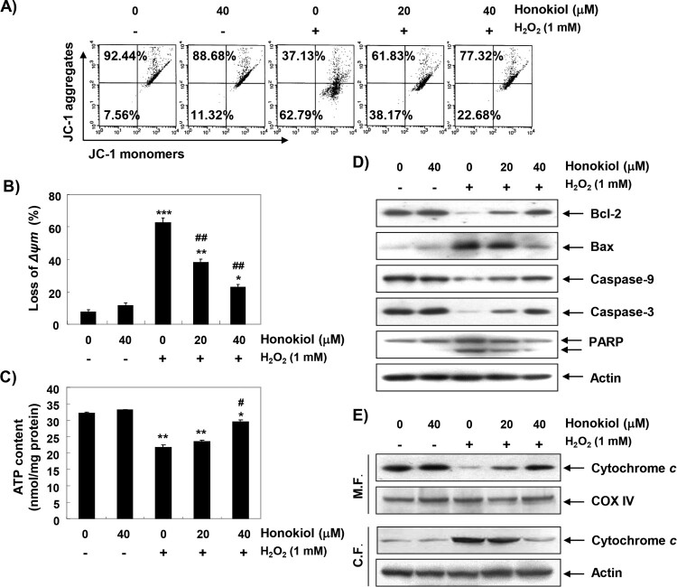
Figure 4.
Attenuation of H2O2-induced mitochondrial dysfunction and changes of apoptosis regulatory proteins by honokiol. Cells were pretreated with honokiol for 1 h, and then stimulated with or without 1 mM H2O2 for 24 h. (A) The cells were incubated with 10 µM JC-1, and the values of MMP were evaluated by a flow cytometer. (B) The data are shown as mean ± SD obtained from three independent experiments. (C) ATP production was monitored using a luminometer. The results are the mean ± SD obtained from three independent experiments. Statistical analyses were conducted using analysis of variance (ANOVA-Tukey’s post hoc test) between groups. *p < 0.05, **p < 0.01 and ***p < 0.001 vs control group, #p < 0.05 and ##p < 0.01 vs H2O2-treated group. (D) The cellular proteins were prepared, and the protein levels were assayed by Western blot analysis. (E) The mitochondrial and cytosolic proteins isolated from cells were separated by SDS polyacrylamide gel electrophoresis, and transferred to the membranes. The membranes were probed with anti-cytochrome c antibody. Equal protein loading was confirmed by the analysis of cytochrome c oxidase subunit IV (COX IV) and actin in each protein extract (M.F., mitochdrial fraction; C.F., cytosolic fraction).

