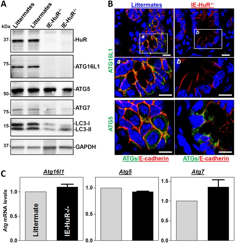
FIG 1.
Targeted deletion of HuR in IECs decreases the levels of intestinal mucosal ATG16L1 in vivo. (A) Immunoblots of HuR and ATGs in the small intestinal mucosae obtained from control littermate and IE-HuR−/− mice. Total proteins were isolated from the intestinal mucosa and prepared for Western blot analysis. Equal loading was monitored by GAPDH. Three experiments were performed and showed similar results. (B) Immunohistochemical staining of ATG16L1 and ATG5 in the small intestinal mucosa. Green, ATG16L1 or ATG5; red, E-cadherin; blue, nuclei stained by DAPI (4′,6′-diamidino-2-phenylindole). Boxes define the area to be amplified. Scale bars, 25 μm. (C) Levels of mRNAs encoding ATG16L1, ATG5, and ATG7 in the small intestinal mucosa as measured by Q-PCR analysis. Values are the means ± SEM (n = 5).

