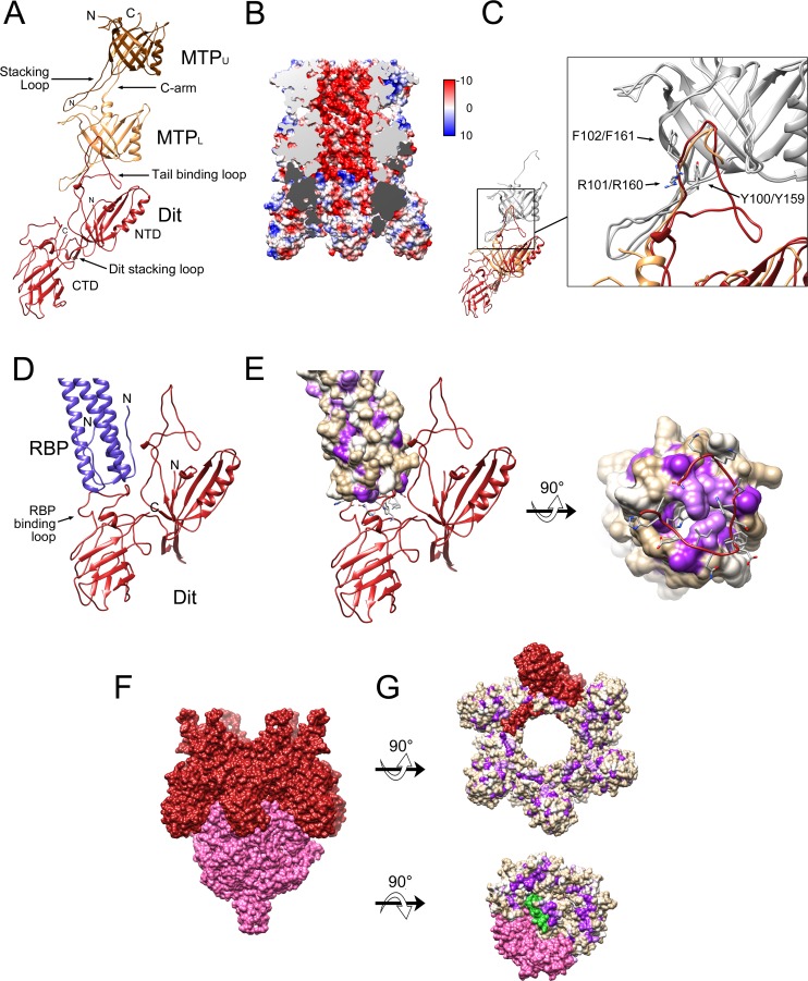
Fig 3. The baseplate core.
(A) One asymmetric unit of the sixfold symmetric baseplate core, showing MTPU (brown), MTPL (tan) and Dit (red). The stacking loops of MTP and Dit, the C-arm of MTPL and the tail binding loop of Dit are indicated. (B) Cutaway electrostatic surface showing the charge distribution inside the tail, colored from red (negative) to blue (positive). The cut surfaces of MTP and Dit are colored light and dark gray, respectively. The surface is colored from –10 (red) to +10 (blue) kcal/(mol*e) according to the color bar. (C) Superposition of MTPU onto MTPL (gray). MTPL (tan) shifted by the same amount then superimposes on Dit (red). The expanded view shows the superposition of the Dit tail binding loop (red) with the MTP C-arm (tan). The side chains of the triplet of residues conserved between the C-arm of MTP (Y159,R160,F161) and the tail binding loop in Dit (Y100,R101,F102) are shown in stick representation and labeled. (D) Detail of the interaction between Dit (red) and the RBP coiled-coil stem region (purple), showing the RBP binding loop in the Dit CTD. (E) Same view as D with RBP shown as a van der Waals surface colored from most hydrophilic (tan) to most hydrophobic (purple), according to the Kyte-Doolittle scale. The rotated view shows the N-terminal end of the RBP trimer and its interaction with the RBP binding loop, with side chains shown in stick representation. (F) Surface representation of Dit (red) and Tal (pink), viewed from the side. (G) Surface representations of Dit (top) and Tal (bottom) rotated 90° in opposite directions to show the interacting surfaces. One subunit each of Dit (red), Tal (pink) and TMP (green) is shown in solid color, the rest are colored by hydrophobicity as in E.

