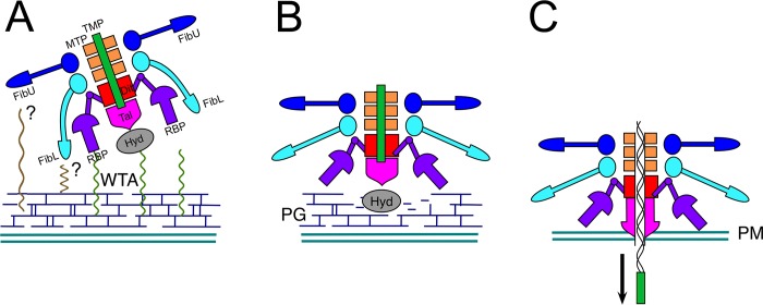
Fig 6. Model for conformational changes in the baseplate during the infection process.
(A) Initial binding of RBP to WTA. FibL and FibU may also be involved in binding to other, as yet unidentified surface structures (?). (B) Conformational changes in RBP lead to exposure of enzymatic activities associated with Hyd and Tal, allowing degradation of the cell wall peptidoglycan (PG). (C) Penetration of the plasma membrane (PM) by the Tal rod helices triggers release of TMP, leading to ejection of DNA through the central channel.

