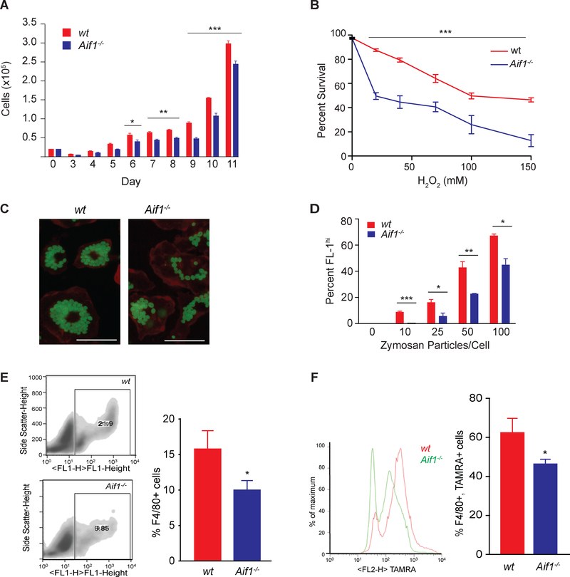
Fig. 2.
Aif1 supports macrophage survival, phagocytosis, and efferocytosis.
(A) Cells were counted during several days after bone marrow harvest using a Coulter counter. (B) Wt and Aif1−/− BMDMs were treated with H2O2 (0, 20, 40, 75, 100 or 150 μM) for 40 minutes. Survival was assessed and quantified with a LIVE/DEAD viability kit. (C) Wt and Aif1−/− BMDMs were synchronized and incubated with the indicated numbers of Alexa 488-zymosan particles per cell (bar, 25 μM). (D) The percentage of internalized zymosan particles per cell was measured by FACS. (E) FACS quantification of the number of F4/80+ cells recruited to the site of inflammation in wt (n=5) or Aif1−/− (n=6) mice. (F) FACS quantification of F4/80+ macrophages isolated from wt (n=5) or Aif1−/− (n=6) that have ingested TAMRA-labeled apoptotic Jurkat T-cells. Error bars indicate ± SEM (* p <0.05, ** p <0.001, *** p <0.0001).

