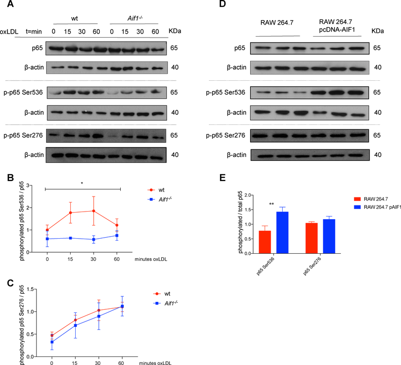
Fig. 3.
Aif1 promotes NF-κB signaling activity.
(A) Wt and Aif1−/− BMDMs were stimulated with oxLDL (50 μg/ml) for 15, 30, or 60 minutes. Lysates were immunoblotted for total p65 and phosphorylated p65 Ser536 and Ser276. (B) Quantification of phosphorylated p65 Ser536 protein levels, relative to total p65 levels. (C) Quantification of phosphorylated p65 Ser276 protein levels, relative to total p65 levels. (D) RAW 264.7 cells were transfected with 2 μg/ml of pcDNA3.1-Aif1 or control pcDNA3.1. 48 hours after transfection lysates were immunoblotted for total p65 and phosphorylated p65 Ser536 and Ser276. (E) Quantification of phosphorylated p65 Ser536 and Ser276 protein levels, relative to total p65 levels. Dashed grey line represents distinct gels. Error bars indicate ± SEM (* p <0.05, ** p <0.001).

