Fig. 5.
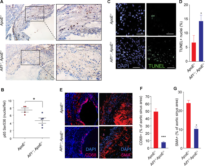
Aif1 promotes NF-κB pathway and limits apoptosis in atherosclerotic plaques in vivo.
(A) Immunohistochemical analysis for phosphorylated p65 Ser536 was performed in atherosclerotic lesions (aortic roots) from Apoe−/− (n=4) and Aif1−/−;ApoE−/− (n=5) mice maintained on high fat diet for 18 weeks. (B) Quantification of phosphorylated p65 Ser536 levels by immunohistochemistry. (C) Representative epifluorescent micrographs showing TUNEL staining of aortic roots from Apoe−/− (n=8) and Aif1−/−;ApoE−/− (n=8) mice (bar, 50 μM) (D) Quantification of TUNEL positive cells on lesions from ApoE−/− or Aif1−/−;ApoE−/− mice. (E) Representative immunofluorescence analysis of CD68+, left, and SMA+, right, cells in aortic roots from Apoe−/− (n= 3) and Aif1−/−;ApoE−/− (n=3) mice maintained on high fat diet for 18 weeks. (F) Quantification of CD68+ cells on lesions from ApoE−/− or Aif1−/−;ApoE−/− mice. (G) Quantification of SMA+ cells on lesions from ApoE−/− or Aif1−/−;ApoE−/− mice. Error bars indicate ± SEM (* p <0.05, *** p <0.0001).

