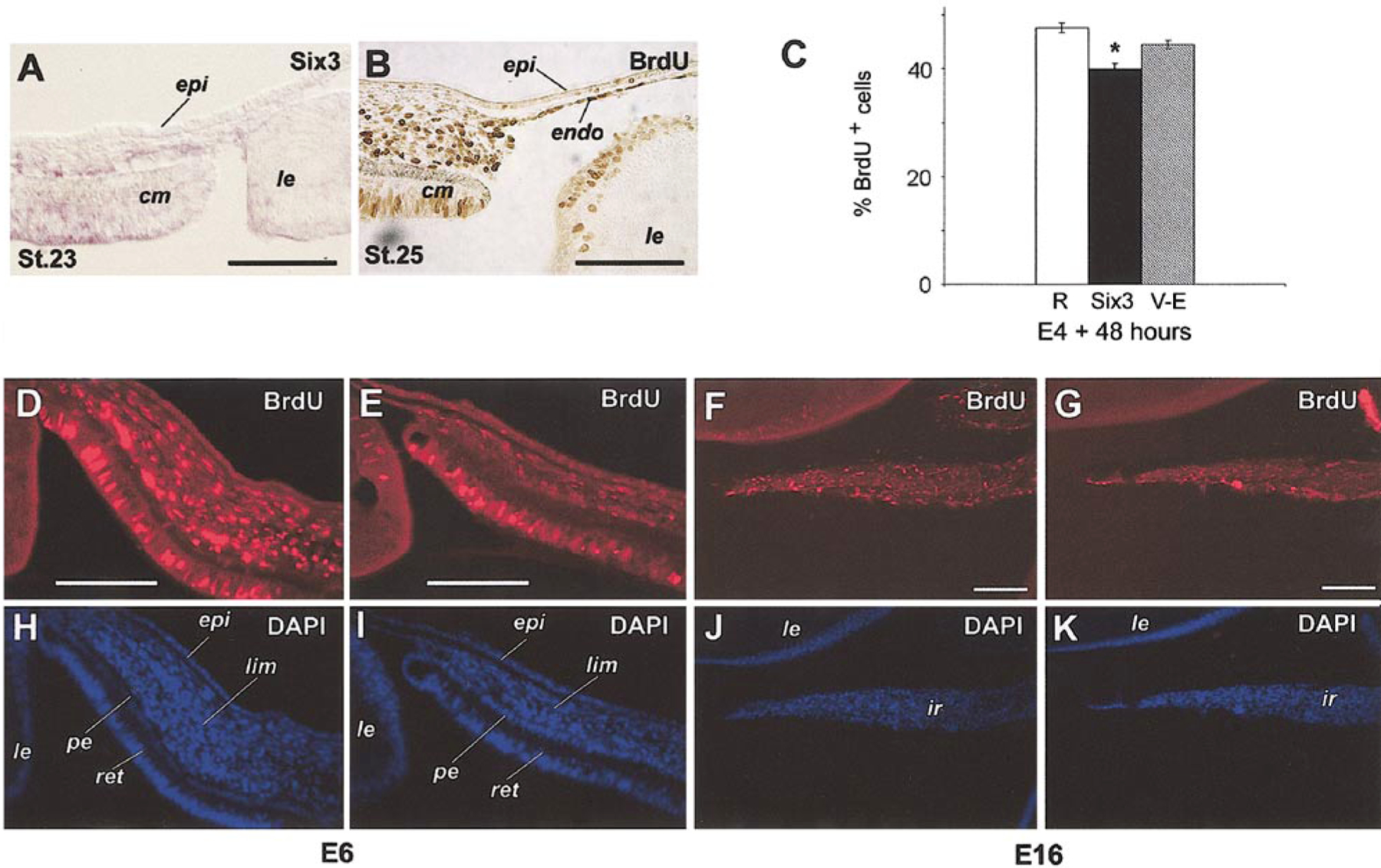
FIG. 8.

Effects of Six3 misexpression on cell proliferation. (A) In situ hybridization of Six3 in the anterior segment at stage 23 (E4). (B) Anti-BrdU immunostaining of eye anterior segment (3 h in vivo labeling) at stage 25 (E5). (C) Quantification of BrdU incorporation in periocular mesenchymal cells in vitro. Percentages of BrdU-positive cells among total cells are shown (average ± standard error). All cells were infected as indicated by coimmunostaining using anti-viral GAG antibody (P27) (data not shown); RCAS virus (R, n = 7), Six3 virus (Six3, n = 7; *, P = 0.015), Six3.V-E virus (V-E, n = 10). (D–G) Anti-BrdU immunostaining compares in vivo BrdU incorporation after infection by Six3 virus (E, G) and RCAS virus (D, F) at stage 10. (D, E) Infected limbal regions at E6 after 3 h of labeling. (F, G) Infected irises at E16 after 6 h of labeling. (H–K) DAPI staining of corresponding fields (D–G), respectively. Extensively viral infection of the same sections was detected by costaining with anti-viral GAG antibody (P27) (data not shown). Scale bars, 100 μm. Abbreviations: epi, corneal epithelium; ir, iris; le, lens; pe, pigmented epithelium; lim, presumptive limbus; ret, retina.
