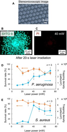
Fig. 2. Fluorescence image and survival rate of high-density LIA on the honeycomb substrate.

(A) Stereomicroscopic image of the honeycomb substrate. (B) Fluorescence image (SYTO 9 staining; green) in a mixed state of live and dead bacteria. (C) Fluorescence image of dead bacteria [propidium iodide (PI) staining; red]. (D) Laser power dependence of P. aeruginosa trapping density and survival rate. (E) Laser power dependence of S. aureus capture density and survival rate.
