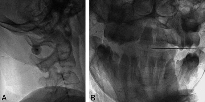
Fig 4.
A, A 14-year-old boy with SMA type 3 and diffuse osseous spinal fusion. With the patient in lateral decubitus position, lateral fluoroscopic image demonstrates the spinal needle superimposed over the C1–C2 interspace. B, The orthogonal view is used for monitoring progress of the needle tip position to midline.

