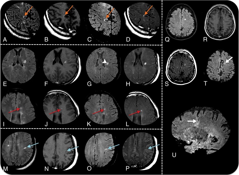
Fig 2.
MR imaging detection of active plaques in 4 different patients with MS. Axial fluid-attenuated inversion recovery sequences (A, E, I, M, and Q), axial Gd-T1 SE sequences (B, F, J, N, and R), axial Gd-SWI sequences (C, G, K, O, and T), T1 Gd-T1 MTC sequences (D, H, L, P, and S), and a sagittal reformatting Gd-SWI sequence (U). Patient 1 demonstrates an acute juxtacortical/cortical demyelinating lesion in the right precuneus with Gd enhancement in all sequences (orange arrows in A–D). Patient 2 demonstrates an acute demyelinating plaque on the left corona radiata (E–F). Gd-SWI depicts the central vein within the plaque (arrowhead in G), adding specificity in favor of the demyelinating substrate. Similar to the Gd-T1 SE and Gd-T1 MTC sequences, the Gd-SWI sequence is also able to demonstrate BBB dysfunction in small lesions (red arrows in I–L). Patient 3 shows an acute demyelinating plaque on the subcortical white matter of the left postcentral gyrus that is only characterized by the Gd-SWI sequence (blue arrows in M–P). Patient 4 has a venous developmental anomaly in the left semiovale center, surrounded by a parenchymal hyperintensity on a FLAIR sequence (Q). Both the Gd-T1 MTC and Gd-SWI sequences (S and T) demonstrate a faint enhancement (arrow in T), which is elegantly demonstrated by the sagittal reformatting Gd-SWI sequence (arrow in U).

