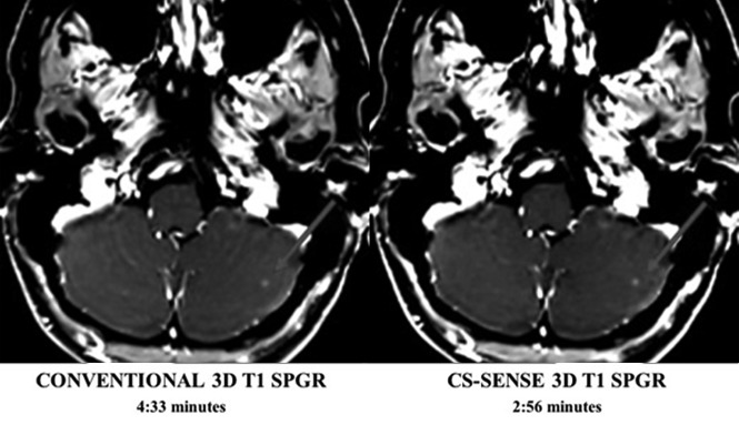
Fig 2.
Conventional and CS-SENSE accelerated axial T1-SPGR images are from the same patient. The arrow demonstrates a small metastasis within the left cerebellar hemisphere that was detected by both sequences equally well. Acquisition of the CS-SENSE SPGR (right) was 35% faster than the conventional SPGR (left).

