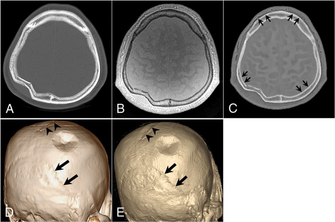
Fig 3.
A 52-year-old man with a right parietal bone fracture. A, Axial CT image. B, Axial proton-density ZTE image. C, Axial CT-like contrast ZTE image. 3D volume-rendered CT image (D) and ZTE skull MR imaging (E). A focal depression fracture is visible in the right parietal bone. The sagittal suture (arrowheads) and bilateral lambdoidal sutures (thick arrows) show conspicuous delineation. Subtle marginal artifacts exist along the inner cortex of both parietal bones and the outer cortices of both frontal bones. The artifacts have a short segmental stepped appearance, which may be related to the postprocessing of histogram-based intensity correction (short thin arrows) in C.

