Figure 3.
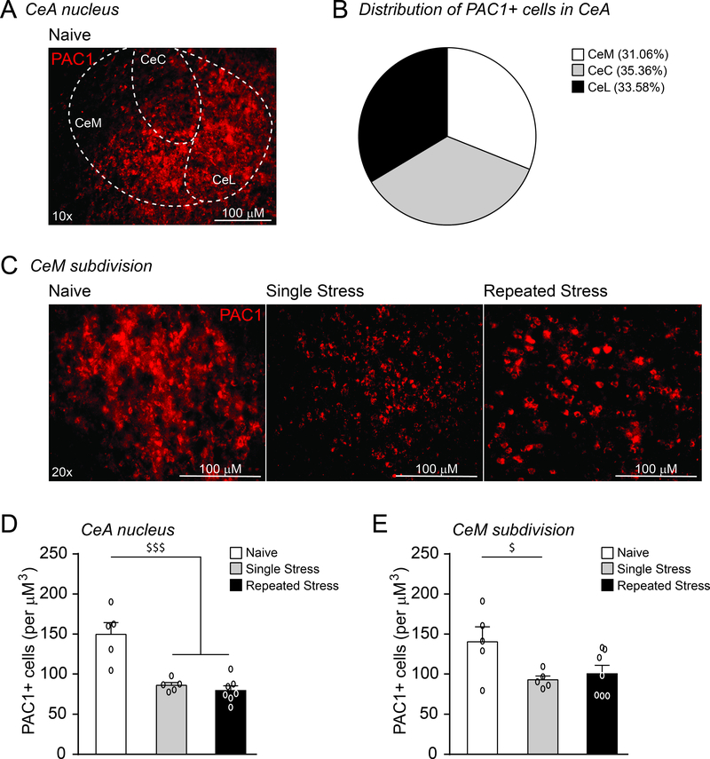
A single restraint stress reduces PAC1 immunoreactivity in the CeM. A. Image of a coronal section through the CeA showing PAC1 immunoreactivity in the lateral (CeL), capsular (CeC) and medial subdivisions (CeM). B. Each CeA subdivision contained a similar number of PAC1+ cells. C. Image of coronal section showing PAC1 immunoreactivity in the CeM of naive rats and rats subjected to single or repeated restraint stress sessions and a 1 hr post-stress recovery period prior to sacrifice. All scale bars represent 100 μM. D. A single restraint period reduced PAC1+ immunoreactivity in the CeA, and this effect persisted with repeated restraint stress sessions (5–7 rats per group). E. A single restraint period reduced the number of PAC1+ cells in the CeM subdivision and a slight recovery was observed followed repeated restraint (5–7 rats per group). All data are presented as mean±SEM. $p<0.05 and $ $ $p<0.001 by one-way ANOVA with post hoc Newman-Keuls multiple comparisons test.

