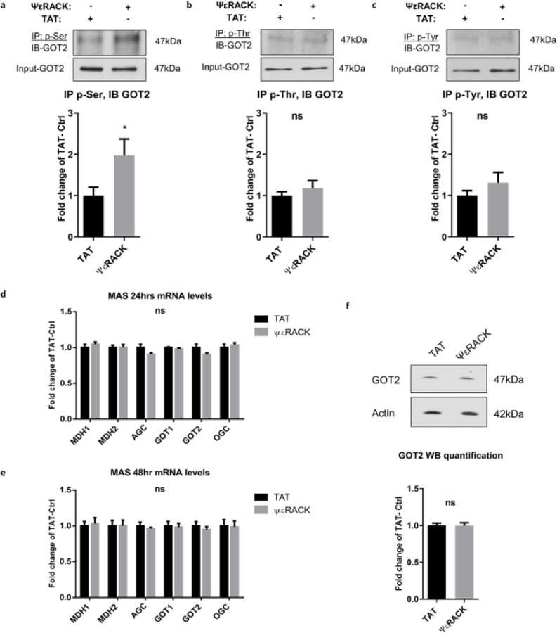
Fig. 3. Effects of PKCε activation on the phosphorylation levels of GOT2 in primary neurons.
(a-c) Neurons were exposed to TAT or ΨεRACK (500 nM) for 1 hour. Forty-eight hours after treatment, protein from cells was immunoprecipitated with (a) phospho-serine, (b) phospho-threonine, (c) phospho-tyrosine, and immunoblotted for GOT2. Input-GOT2 represents as a loading control. Quantification fold change of TAT control and representative blot as shown above. (d-f) PKCε activation does not alter the expression of MAS components in neurons. Real-time qPCR was performed (d) 24 hours and (e) 48 hours following the 1-hour treatment of TAT or ΨεRACK (500 nM) in primary neurons. mRNA levels were normalized to GAPDH and shown as fold change of TAT control. (f) Western blots of GOT2 48 hours following the 1-hour treatment with TAT or ΨεRACK (500 nM) in neurons. Actin acts as a loading control. Quantification fold change of TAT control and representative blot as shown above (n=5–7, mean ± SEM, * P <0.05, ns = non-significant, Two-tailed unpaired Student’s t-test). MDH1: Malate dehydrogenase 1; MDH2: Malate dehydrogenase 2; AGC: Aspartate glutamate carrier; GOT1: Glutamic oxaloacetic transaminase 1; GOT2: Glutamic oxaloacetic transaminase 2; OGC: Oxoglutarate carrier.

