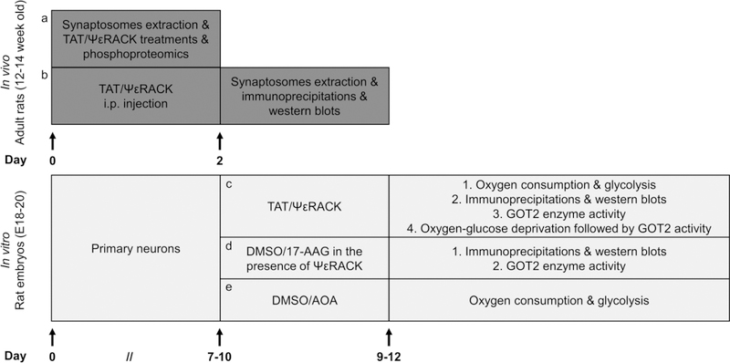
Fig. 7. Schematic diagram of the experimental design.
Briefly, (a, b) synaptosomes were extracted from adult rats. Phosphoproteomic analysis, immunoprecipitation and immunoblots were used to examine PKCε-activated phosphorylation. (c-e) Primary neurons were prepared from embryonic 18–20 day old pups. Oxygen consumption and glycolysis were evaluated by Seahorse Bioscience Technology. PKCε-mediated phosphorylation was determined by immunoprecipitation and western blots. A colorimetric assay was utilized to assess the GOT2 activity. We used oxygen-glucose deprivation as an in vitro model to evaluate the effects of ischemic injury on GOT2 activity. For pharmacological treatments and experimental details please refer to “Materials and Methods”.

