
Figure 3.
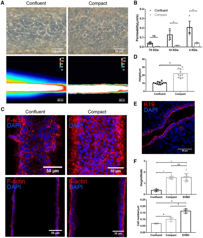
Cholangiocyte monolayers require high confluency for mature barrier function. (A) Representative bright‐field images (upper panels) and pseudo color images after FITC‐dextran (4 kDa) perfusion for 2 minutes (lower panels) of confluent and compact cholangiocyte channels. Scale bar: 100 μm (upper panels), 200 μm (lower panels). (B) Permeability of confluent and compact cholangiocyte channel to FITC‐dextran (70, 10, and 4 kDa), n = 8 devices. (C) Bottom (upper panels) and middle (lower panels) views of confluent and compact cholangiocyte monolayers in the devices, stained for F‐actin (red) and nuclei (DAPI; blue). Scale bars, 50 μm. (D) Cell height of confluent and compact cholangiocyte monolayers in the devices, n ≥ 10. (E) Adult mouse extrahepatic bile duct, stained for K19 (red) and nuclei (DAPI; blue), representative images from n = 9. Scale bar, 50 μm. (F) Cell height/width ratio (upper panel) and cell density (lower panel) in confluent and compact cholangiocyte channels and mice EHBD, n = 4‐9. Images are representative of at least three independent experiments. All data are presented as mean ± SD; *P < 0.05.