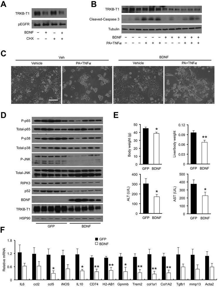
Figure 7. BDNF decreases membrane TRKB-T1 and protects mice from diet-induced NASH.
(A) Immunoblots of membrane proteins isolated from Hepa 1–6 cells stably expressing TrkB-T1. Transduced cells were treated with vehicle (−) or BDNF in the presence or absence of cycloheximide (CHX; 4 μM).
(B) Immunoblots of total cell lysates from primary hepatocytes isolated from hnRNPU LKO mice. Hepatocytes were pretreated with BDNF (100 ng/mL) for 2 hrs before addition of PA (200 μM)) and TNFa (20 ng/mL).
(C) Morphology of treated hepatocytes. Hepatocytes were pretreated with BDNF (100 ng/mL) for 2 hrs before addition of PA (400 μM) and TNFa (20 ng/mL).
(D) Immunoblots of total liver lysates from mice transduced with AAV-GFP or AAV-BDNF (GFP n=7; BDNF n=6).
(E) Metabolic parameters and plasma ALT/AST levels in transduced mice.
(F) qPCR analysis of hepatic gene expression. Data in (E) and (F) represent mean ± SEM. *P<0.05, **P<0.01; BDNF vs. GFP, two-tailed unpaired Student’s t-test.

