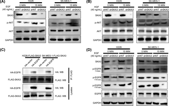
Figure 4. SKA3 and EGFR interact to enhance PI3K–AKT signaling.
(A and B) Cells were stimulated with EGF (A) or HGF (B) for 0, 5 or 15 min and assessed for the expression of the indicated proteins. GAPDH was probed as a loading control. (C) Co-Ips using anti-FLAG antibodies in LUAD cells co-transfected with FLAG-SKA3 along with HA-EGFR or empty vector controls, as specified. (D) Cells were EGF treated for 0 or 5 min.

