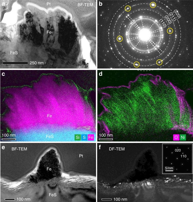
Fig. 2. Detailed structures of metallic iron whiskers on troilite.
a A TEM bright-field (BF) image of a whisker on troilite in a FIB section. b A SAED pattern of the whole region of the whisker. Dotted rings have radii corresponding to the d-spacings of {hkl}planes of bcc iron. Pairs of diffraction spots from the same crystals are connected by dotted lines. The six circled spots are reflected from a single iron crystal belonging to a zone axis [1 ®20]. c-d X-ray intensity maps of the whisker in (a). Composite image of Si (green), S (cyan), and Fe (magenta) is shown in (c) and that of O (magenta) and Ni (green) is displayed in (d). e A TEM-BF image of a thinner whisker in a FIB section from an edge-on view (the electron beam was nearly parallel to the troilite surface). f A TEM-dark field (DF) image corresponding to (e). A SAED pattern obtained from the whisker is shown on the upper right, showing bcc iron in zone axis [001]. a-d was obtained from FIB section RA-QD02-0292-01 (Supplementary Fig. 2). e-f was obtained from FIB section RA-QD02-0325-01 (Supplementary Fig. 3).

