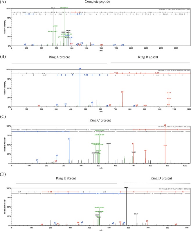
Figure 6.
MS/MS spectra for nisin P peptides obtained by trypsin digestion and generated in Scaffold (v4.4.1.1, www.proteomsoftware.com). b- ions are represented in red and y- ions in blue. (A) Spectrum of nisin P showing fragmentation only in linear region, indicating presence of all rings. (B) Spectrum showing presence of ring A (C is free and there is no fragmentation) and absence of ring B (C has a CAM modification and is fragmented). (C) detail of presence of ring C and hinge region, AIK (D). Presence of ring D and CAM modification indicating the absence of ring E.

