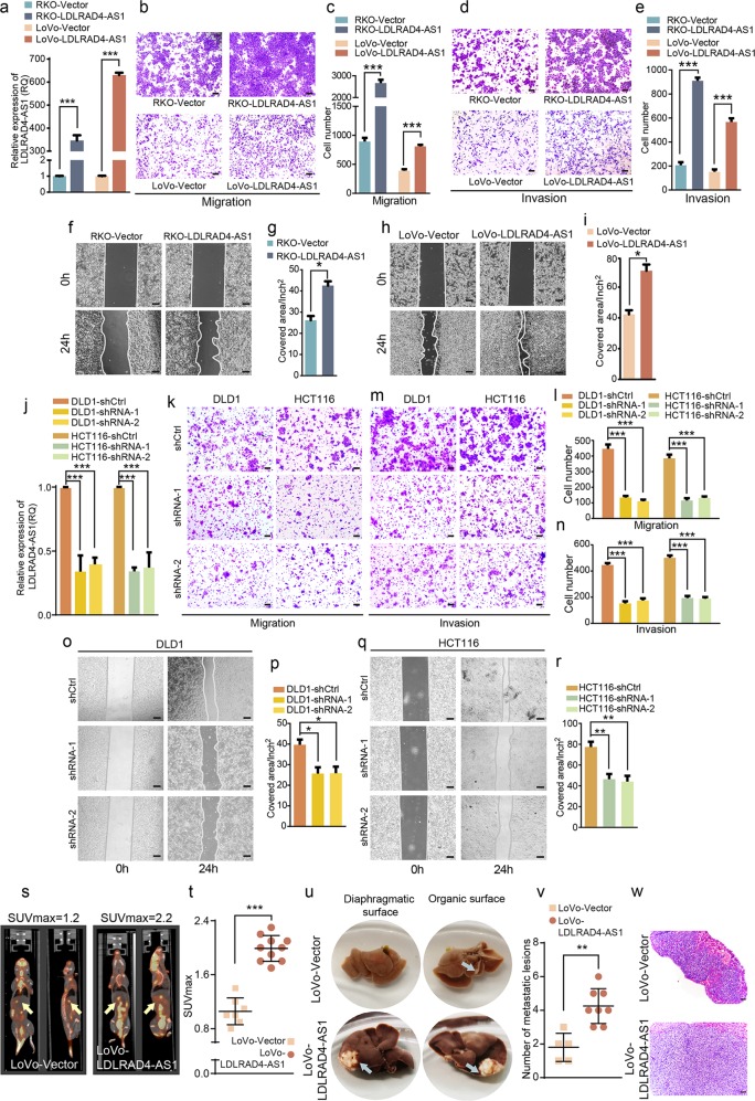
Fig. 2. LncRNA LDLRAD4-AS1 promotes CRC cell migration and invasion in vitro as well as promotes CRC metastasis in vivo.
a LDLRAD4-AS1-overexpressing RKO and LoVo cell lines were established by the transfection of pCDH- LDLRAD4-AS1. LncRNA LDLRAD4-AS1 levels in cells were detected by qRT-PCR. b, c Migration assays were used to determine the effects of LDLRAD4-AS1 overexpression on the migration ability of CRC cells. d, e Invasion assays were used to determine the effects of LDLRAD4-AS1 overexpression on the invasion ability of CRC cells. f–i The migration potencies of CRC cells with the indicated treatments were detected by using wound-healing assay. j infecting DLD1 and HCT116 cells with a lentivirus vector harboring shRNA-LDLRAD4-AS1 was to knock down the endogenous expression of lncRNA LDLRAD4-AS1 in cells. lncRNA LDLRAD4-AS1 levels in cells were detected by qRT-PCR. k, l Migration assays were used to determine the effects of LDLRAD4-AS1-depleted on the migration ability of CRC cells. m, n Invasion assays were used to determine the effects of LDLRAD4-AS1-depleted on the invasion ability of CRC cells. o–r The migration potencies of CRC cells with the indicated treatments were detected by using wound-healing assay. s Representative photographs of 18F-FDG PET/CT scans of LoVo-LDLRAD4-AS1 and control LoVo-Vector-injected mice. t The SUVmax was higher in the LoVo-LDLRAD4-AS1 group than in the control group. u The gross images of liver metastases observed in the nude mice injected with LoVo cells. Arrows represent metastatic tumors. v The number of metastatic lesions was larger in the LoVo-LDLRAD4-AS1 group than in the control group. w H&E staining for the liver metastases. For a–w, data were expressed as means ± SD in three independent experiments. *p < 0.05, **p < 0.01, ***p < 0.001.

