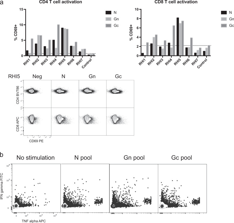
Fig. 3. Functional assays of RVFV-specific T cells in vaccine recipients.
a In vitro exposure of PBMCs from most vaccine recipients to the down-selected panel of peptides (Table 2) for each structural protein resulted in upregulation of the T cell activation marker CD69 on both CD4 and CD8 T cells. The dotted line shows the background frequency based upon the highest frequency of expression observed in the negative control. Representative raw flow plots depicting CD69 expression are shown for the donor with the highest RVFV-specific T cell activity (RHI5). b PBMCs from RHI5 also expressed IFN-γ and TNF-α following peptide stimulation.

